Publications Ullrich Lab
The ultrafast internal conversion and intersystem crossing dynamics of 2-thiouracil (2TU) and 2-thiothymine (2TT) are studied using time-resolved photoelectron spectroscopy to investigate the effect of methylation on the deactivation mechanism. Like other thiobases, the triplet manifold is populated with high quantum yields via the lowest singlet excited state, which is dark in absorption. This study focuses on the lowest triplet state and the role of two minima, with sulfur-out-of-plane and slightly boat-like geometries, in the intersystem crossing dynamics back to the ground state.
The ultrafast internal conversion (IC) and intersystem crossing (ISC) dynamics of 2-thiouracil (2TU) and 2-thiothymine (2TT) are studied with time-resolved photoelectron spectroscopy (TRPES) to investigate the effect of methylation on the deactivation mechanism.
Porphyrins play pivotal roles in many crucial biological processes including photosynthesis. However, there is still a knowledge gap in understanding electronic and excited state implications associated with functionalization of the porphyrin ring system. These effects can have electrochemical and spectroscopic signatures that reveal the complex nature of these somewhat minor substitutions, beyond simple inductive or electronic effect correlations. To obtain a deeper insight into the influences of porphyrin functionalization, four free-base, meso-substituted porphyrins: tetraphenyl porphyrin (TPP), tetra(4-hydroxyphenyl) porphyrin (THPP), tetra(4-carboxyphenyl) porphyrin (TCPP), and tetra(4-nitrophenyl) porphyrin (TNPP), were synthesized, characterized, and investigated. The influence of various substituents, (-hydroxy,-carboxy, and -nitro) in the para position of the meso-substituted phenyl moieties were evaluated by spectroelectrochemical techniques (absorption and fluorescence), femtosecond transient absorption spectroscopy, cyclic and differential pulse voltammetry, ultraviolet photoelectron spectroscopy (UPS), and time-dependent density functional theory (TD-DFT). Spectral features were evaluated for the neutral porphyrins and differences observed among the various porphyrins were further explained using rendered frontier molecular orbitals pertaining to the relevant transitions. Electrochemically generated anionic and cationic porphyrin species indicate similar absorbance spectroscopic signatures attributed to a red-shift in the Soret band. Emissive behavior reveals the emergence of one new fluorescence decay pathway for the ionic porphyrin, distinct from the neutral macrocycle. Femtosecond transient absorption spectroscopy analysis provided further analysis of the implications on the excited-state as a function of the para substituent of the free-base meso-substituted tetraphenyl porphyrins. Herein, we provide an in-depth and comprehensive analysis of the electronic and excited state effects associated with systematically varying the induced dipole at the methine bridge of the free-base porphyrin macrocycle and the spectroscopic signatures related to the neutral, anionic, and cationic species of these porphyrins.
Photo-oxa-dibenzocyclooctyne (Photo-ODIBO) undergoes photodecarbonylation under UV excitation to its bright S2 state, forming a highly reactive cyclooctyne, ODIBO. Following 321 nm excitation with sub-50 fs actinic pulses, the excited state evolution and cyclopropenone bond cleavage with CO release were characterized using femtosecond stimulated Raman spectroscopy and time-dependent density functional theory Raman calculations. Analysis of the photo-ODIBO S2 C=O Raman band revealed multi-exponential intensity, peak splitting and frequency-shift dynamics. This suggests a stepwise cleavage of the two C–C bonds in the cyclopropenone structure that is completed within less than 300 fs after excitation. Evidence of intramolecular vibrational relaxation on the S2 state, concurrent with photodecarbonylation, with dynamics matching previous electronic transient absorption spectroscopy, was also observed. This confirms an excited state, as opposed to ground state, photodecarbonylation mechanism resulting in a vibronically excited photoproduct, ODIBO.
The ultrafast dynamics of photo-OxaDiBenzocycloOctyne (photo-ODIBO) photo-dissociation was studied using femtosecond transient absorption spectroscopy. Steady-state UV–Vis, time-dependent density functional theory, and 350 nm and 321 nm transient absorption studies are reported. Photo-ODIBO excitation with 321 nm and 350 nm light-induced photodecarbonylation of the cyclopropenone functional group results in the formation of ODIBO. The presence of the photoproduct was confirmed by the results of steady-state photolysis experiments and the observation of absorption signatures of ODIBO in the photo-ODIBO transient absorption spectra. Analysis of the latter revealed the underlying photochemical mechanisms and associated time constants, following excitation of the samples. The dynamics show a multi-exponential decay process, following the dissociation of photo-ODIBO into an excited state of the photoproduct ODIBO within <294 fs after 321 nm excitation. 350 nm excitation, on the other hand, is shown to produce ground state ODIBO via an intermediate species. Additional transient absorption measurements were performed directly on the photoproduct ODIBO to help distinguish spectral signatures associated with these processes.
Cu2O, CuO, and mixed-phase Cu2O/CuO thin films with different relative compositions were prepared by oxidizing Cu films at temperatures 150–380 °C for a time period ranging from 2 to 24 h, and their ultrafast transient absorption spectra have been characterized to understand the carrier dynamics of the heterostructured Cu2O/CuO system. The absorption dynamics of a pure p-type Cu2O sample followed a biexponential decay, with a fast time ∼0.3 ps and a long life >150 ps, while a pure p-type CuO sample showed triexponential decay dynamics, with three time constants, 0.25, 2.5, and >150 ps. For the mixed-phased Cu2O/CuO thin films, their absorption dynamics all followed the triexponential decay, and the two ultrafast time constants showed strong composition dependence. Possible energy band structures and electron transition processes are proposed to understand both the spectroscopic and dynamics behaviors of these samples.
The photophysical properties of 2,4-dithiouracil (2,4-DTU) in the gas phase are studied by time-resolved photoelectron spectroscopy (TRPES) with three different excitation wavelengths in direct extension of previous work on uracil (U), 2-thiouracil (2-TU) and 4-thiouracil (4-TU). Non-radiative deactivation in the canonical nucleobases like uracil mainly occurs via internal conversion (IC) along singlet excited states, although intersystem crossing (ISC) to a long-lived triplet state was confirmed to play a minor role. In thionated uracils, ISC to the triplet state becomes ultrafast and highly efficient with a quantum yield near unity; however, the lifetime of the triplet state is strongly dependent on the position of the sulfur atom. In 2-TU, ISC back to the ground state occurs within a few hundred picoseconds, whereas the population remains trapped in the lowest triplet state in the case of 4-TU. Upon doubling the degree of thionation, ISC remains highly efficient and dominates the photophysics of 2,4-DTU. However, several low-lying excited states contribute to competing IC and ISC pathways and a complex deactivation mechanism, which is evaluated here based on TRPES measurements and discussed in the context of the singly thionated uracils.
SPOC (SPectral Ocean Color) is a 3U small satellite mission that will use an adjustable multispectral imager to map sensitive coastal regions and off coast water quality of Georgia and beyond. SPOC is being developed by the University of Georgia’s (UGA) Small Satellite Research Laboratory (SSRL) through NASA’s Undergraduate Student Instrument Project (USIP). UGA is working with Cloudland Instruments to develop a small scale (<1000 cm3) multispectral imager, ranging from 400-850nm, for Earth science applications which will fly as part of the NASA CubeSat Launch Initiative.The project is UGA’s first satellite mission and is built by a team of undergraduates from a wide range of backgrounds and supervised by a multidisciplinary team of graduate students and faculty. Development, assembly, testing, and validation of the multispectral imager, as well integrating it into the satellite are all being done in house. At an orbit of 400 km the resulting images will be 90 km x 100 km in size, with a default spatial resolution and spectral resolution of 130 m and 4 nm, respectively.
The photophysics of thionated uracils are investigated using time-resolved photoelectron spectroscopy with emphasis on evaluating differences in intersystem crossing dynamics with respect to substituent position.
Femtosecond time-resolved spectroscopy has been used to study the light-induced bioenergetics in isolated cyanobacterial photosystem I (PSI) reaction centers from Synechocystis sp. PCC 6803 at 77K. A method was developed to produce optically clear samples at 77 K without the use of cryprotectants, which considerably simplifies the experiments. Relatively intense laser pulses were used for sample excitation. Following 400 nm excitation, predominantly chlorophyll a pigments in PSI with Qy absorption maxima in the 670–685 nm region are excited. This initially excited distribution of pigments transfers energy in ~400 fs to pigments absorbing near 690 nm, and to pigments absorbing near 715 nm. Further equilibration processes occur and are characterized by a 2.8 ps time constant. Following equilibration, energy trapping and formation of the secondary radical pair state, P700+A1–, occurs with a time constant of 32.5 ps
We report time-dependent photoelectron spectra recorded with a single-photon ionization setup and extensive simulations of the same spectra for the excited-state dynamics of 2-thiouracil (2TU) in the gas phase. We find that single-photon ionization produces very similar results as two-photon ionization, showing that the probe process does not have a strong influence on the measured dynamics. The good agreement between the single-photon ionization experiments and the simulations shows that the norms of Dyson orbitals allow for qualitatively describing the ionization probabilities of 2TU. This reasonable performance of Dyson norms is attributed to the particular electronic structure of 2TU, where all important neutral and ionic states involve similar orbital transitions and thus the shape of the Dyson orbitals do not strongly depend on the initial neutral and final ionic state. We argue that similar situations should also occur in other biologically relevant thio-nucleobases, and that the time-resolved photoelectron spectra of these bases could therefore be adequately modeled with the techniques employed here.
The ultraviolet (UV) photophysics of the natural and modified nucleobases can be surprisingly different. In response to UV radiation, the natural pyrimidine nucleobases undergo ultrafast internal conversion back to the ground state, whereas their thiobase analogues, in which an oxygen has been replaced by sulfur, instead display efficient intersystem crossing to the triplet manifold. Here, the effect of the substituent position is investigated with time-resolved photoelectron spectroscopy on 4-thiouracil, which is contrasted to previous work on 2-thiouracil. Although the photophysical pathway of both structural isomers is similar, i.e., leading from the S2 (ππ*) state, via S1 (nπ*), to the triplet manifold and subsequently back to the ground state, the intersystem crossing dynamics are strongly influenced by the surrounding intramolecular environment of the sulfur atom.
Irradiation of cyclopropenone-masked dibenzocyclooctynes with near-infrared pulses from a femtosecond laser triggers photodecarbonylation via nonresonant two- or three-photon excitation. Multiphoton-generated cyclooctynes undergo a SPAAC reaction with organic azides, yielding the expected triazoles. Multiphoton-triggered SPAAC (MP-SPAAC) enables high resolution 3-D photoclick derivatization of hydrogels and tissues.
Single-atom substitution within a natural nucleobase—such as replacing oxygen by sulfur in uracil—can result in drastic changes in the relaxation dynamics after UV excitation. While the photodynamics of natural nucleobases like uracil are dominated by pathways along singlet excited states, the photodynamics of thiobases like 2-thiouracil populate the triplet manifold with near unity quantum yield. In the present study, a synergistic approach based on time-resolved photoelectron spectroscopy (TRPES), time-resolved absorption spectroscopy (TRAS), and ab initio computations has been particularly successful at unraveling the underlying photophysical principles and describing the dissimilarities between the natural and substituted nucleobases. Specifically, we find that varying the excitation wavelength leads to differences between gas-phase and condensed-phase experimental results. Systematic trends are observed in the intersystem crossing time constants with varying excitation wavelength, which can be readily interpreted in the context of ab initio calculations performed both in vacuum and including solvent effects. Thus, the combination of TRPES and TRAS experiments with high-level computational techniques allows us to characterize the topology of the potential energy surfaces defining the relaxation dynamics of 2-thiouracil in both gas and condensed phases, as well as investigate the accessibility of conical intersections and crossings, and potential energy barriers along the associated relaxation coordinates.
The photophysics of uracil and 2-thiouracil have been investigated using time-resolved photoelectron spectroscopy with emphasis on evaluating the role of intersystem crossing pathways.
The photodynamic properties of molecules determine their ability to survive in harsh radiation environments. As such, the photostability of heterocyclic aromatic compounds to electromagnetic radiation is expected to have been one of the selection pressures influencing the prebiotic chemistry on early Earth. In the present study, the gas-phase photodynamics of uracil, 5-methyluracil (thymine) and 2-thiouracil—three heterocyclic compounds thought to be present during this era—are assessed in the context of their recently proposed intersystem crossing pathways that compete with internal conversion to the ground state. Specifically, timeresolved photoelectron spectroscopy measurements evidence femtosecond to picosecond timescales for relaxation of the singlet 1pp* and1np* states as well as for intersystem crossing to the triplet manifold. Trapping in the excited triplet state and intersystem crossing back to the ground state are investigated as potential factors contributing to the susceptibility of these molecules to ultraviolet photodamage.
A wavelength dependent study investigating the low-lying 1La and 1Lb states, both possessing 1ππ* character, and the 1πσ* state in the deactivation process of indole is presented here. Relaxation dynamics following excitation at 241, 250, 260, 270, 273, and 282 nm are examined using three gas-phase, pump-probe spectroscopic techniques: (1) hydrogen atom (H-atom) time-resolved kinetic energy release (TR-KER), (2) time-resolved photoelectron spectroscopy (TR-PES), and (3) time-resolved ion yield (TR-IY). Applied in combination, a more complete picture of the indole relaxation dynamics may be gleaned. For instance, TR-PES experiments directly observe all relaxation pathways by probing the evolution of the excited states following photoexcitation; whereas, TR-KER measurements indirectly, yet specifically, probe for 1πσ*-state activity through the detection of H-atoms eliminated along the indole nitrogen-hydrogen (N-H) stretch coordinate—a possible outcome of 1πσ*-state relaxation in indole. In addition, mass information obtained via TR-IY monitors fragmentation dynamics that may occur within the neutral electronically excited and/or cationic states. The work herein assesses the onset and importance of the 1πσ* state at various pump wavelengths by systematically tuning across the ultraviolet absorption spectrum of indole with a particular focus on those pump wavelengths longer than 263 nm, where the involvement of the 1πσ* state is under current debate. As far as this experimental work is concerned, there does not appear to be any significant involvement by the 1πσ* state in the indole relaxation processes following excitation at 270, 273, or 282 nm. This investigation also evaluates the primary orbital promotions contributing to the 1La, 1Lb, and 1πσ* transitions based on ionization preferences observed in TR-PES spectra. Relaxation time constants associated with dynamics along these states are also reported for excitation at all of the aforementioned pump wavelengths and are used to pinpoint the origin of the discrepancies found in the literature. In this context, advantages and disadvantages of the three experimental techniques are discussed.
We have investigated the effects of quantum tunneling on the photodissociation dynamics of ammonia, following below and above barrier photoexcitation of low-lying levels of the ν2′ umbrella mode of the NH3 à state (NH3 (Ã)). This barrier separates the local minimum of the vertical Franck–Condon region from the NH3 (Ã)/NH3 (X̃) conical intersection (CI) which can be accessed along the N–H stretch coordinate. Two complementary techniques, time-resolved photoelectron spectroscopy (TR-PES) and time-resolved total kinetic energy release spectroscopy (TR-TKER), have been utilized to directly measure, for the first time, vibrational level dependent excited state lifetimes and N–H dissociation time scales as well as photoproduct final energy distributions. Interestingly, ν2′ even/odd dependencies are observed in the measured time constants and NH2 internal energy spectra, which are attributed to tunneling through a barrier, whose magnitude is dependent on the planarity of NH3 in the à state and direct versus indirect dissociation at the NH3 (Ã)/NH3 (X̃) conical intersection.
The studies herein investigate the involvement of the low-lying 1La and 1Lb states with 1ππ* character and the 1πσ* state in the deactivation process of indole following photoexcitation at 201 nm. Three gas-phase, pump-probe spectroscopic techniques are employed: (1) Time-resolved photoelectron spectroscopy (TR-PES), (2) hydrogen atom (H-atom) time-resolved kinetic energy release (TR-KER), and (3) time-resolved ion yield (TR-IY). Each technique provides complementary information specific to the photophysical processes in the indole molecule. In conjunction, a thorough examination of the electronically excited states in the relaxation process, with particular focus on the involvement of the 1πσ* state, is afforded. Through an extensive analysis of the TR-PES data presented here, it is deduced that the initial excitation of the 1Bb state decays to the 1La state on a timescale beyond the resolution of the current experimental setup. Relaxation proceeds on the 1La state with an ultrafast decay constant (<100 femtoseconds (fs)) to the lower-lying 1Lb state, which is found to possess a relatively long lifetime of 23 ± 5 picoseconds (ps) before regressing to the ground state. These studies also manifest an additional component with a relaxation time of 405 ± 76 fs, which is correlated with activity along the 1πσ* state. TR-KER and TR-IY experiments, both specifically probing 1πσ* dynamics, exhibit similar decay constants, further validating these observations.
The heteroaromatic ultraviolet chromophore pyrrole is found as a subunit in a number of important biomolecules: it is present in heme, the non-protein component of hemoglobin, and in the amino acid tryptophan. To date there have been several experimental studies, in both the time- and frequency-domains, which have interrogated the excited state dynamics of pyrrole. In this work, we specifically aim to unravel any differences in the H-atom elimination dynamics from pyrrole across an excitation wavelength range of 250–200 nm, which encompasses: (i) direct excitation to the (formally electric dipole forbidden) 11πσ* (1A2) state; and (ii) initial photoexcitation to the higher energy 1ππ* (1B2) state. This is achieved by using a combination of ultrafast time-resolved ion yield and time-resolved velocity map ion imaging techniques in the gas phase. Following direct excitation to 11πσ* (1A2) at 250 nm, we observe a single time-constant of 126 ± 28 fs for N–H bond fission. We assign this to tunnelling out of the quasi-bound 3s Rydberg component of the 11πσ* (1A2) surface in the vertical Franck–Condon region, followed by non-adiabatic coupling through a 11πσ*/S0 conical intersection to yield pyrrolyl radicals in their electronic ground state (C4H4N([X with combining tilde])) together with H-atoms. At 238 nm, direct excitation to, and N–H dissociation along, the 11πσ* (1A2) surface is observed to occur with a time-constant of 46 ± 22 fs. Upon initial population of the 1ππ* (1B2) state at 200 nm, a rapid 1ππ* (1B2) → 11πσ* (1A2) → N–H fission process takes place within 52 ± 12 fs. In addition to ultrafast N–H bond cleavage at 200 nm, we also observe the onset of statistical unimolecular H-atom elimination from vibrationally hot S0 ground state species, formed after the relaxation of excited electronic states, with a time-constant of 1.0 ± 0.4 ns. Analogous measurements on pyrrole-d1 reveal that these statistical H-atoms are released only through C–H bond cleavage.
Time-resolved photoelectron spectra of ammonia display combination bands of the umbrella and stretching modes associated with the N-H coordinate of s* relaxation. Time-resolved photodissociation studies determine timescales less than 200fs. Similar s* photochemistry is found in heteroaromatics
A combination of ultrafast time-resolved velocity map imaging (TR-VMI) methods and complete active space self-consistent field (CASSCF) ab initio calculations are implemented to investigate the electronic excited-state dynamics in aniline (aminobenzene), with a perspective for modeling 1πσ* mediated dynamics along the amino moiety in the purine derived DNA bases. This synergy between experiment and theory has enabled a comprehensive picture of the photochemical pathways/conical intersections (CIs), which govern the dynamics in aniline, to be established over a wide range of excitation wavelengths. TR-VMI studies following excitation to the lowest-lying 1ππ* state (11ππ*) with a broadband femtosecond laser pulse, centered at wavelengths longer than 250 nm (4.97 eV), do not generate any measurable signature for 1πσ* driven N–H bond fission on the amino group. Between wavelengths of 250 and greater than 240 nm (less than 5.17 eV), coupling from 11ππ* onto the 1πσ* state at a 11ππ*/1πσ* CI facilitates ultrafast nonadiabatic N–H bond fission through a 1πσ*/S0 CI in less than 1 ps, a notion supported by CASSCF results. For excitation to the higher lying 21ππ* state, calculations reveal a near barrierless pathway for CI coupling between the 21ππ* and 11ππ* states, enabling the excited-state population to evolve through a rapid sequential 21ππ* → 11ππ* → 1πσ* → N–H fission mechanism, which we observe to take place in 155 ± 30 fs at 240 nm. We also postulate that an analogous cascade of CI couplings facilitates N–H bond scission along the 1πσ* state in 170 ± 20 fs, following 200 nm (6.21 eV) excitation to the 31ππ* surface. Particularly illuminating is the fact that a number of the CASSCF calculated CI geometries in aniline bear an exceptional resemblance with previously calculated CIs and potential energy profiles along the amino moiety in guanine, strongly suggesting that the results here may act as an excellent grounding for better understanding 1πσ* driven dynamics in this ubiquitous genetic building block.
The ultrafast excited state relaxation of ammonia is investigated by resonantly exciting specific vibrational modes of the electronically excited NH3 (Ã) state using three complementary femtosecond (fs) pump–probe techniques: time-resolved photoelectron, ion-yield and photofragment translational spectroscopy. Ammonia can be seen as a prototypical system for studying non-adiabatic dynamics and therefore offers a benchmark species for demonstrating the advantages of combining the aforementioned techniques to probe excited state dynamics, whilst simultaneously illuminating new aspects of ammonia's photochemistry. Time-resolved photoelectron spectroscopy (TRPES) provides direct spectroscopic evidence of σ* mediated relaxation of the NH3 (Ã) state which manifests itself as coupling of the umbrella (ν2) and symmetric N–H stretch (ν1) modes in the photoelectron spectra. Time-resolved ion yield (TRIY) and time-resolved photofragment translation spectroscopy (TRPTS) grant a measure of the dissociation dynamics through analysis of the H and NH2 photodissociation co-fragments. Initial vibrational level dependent TRIY measurements reveal photoproduct formation times of between 190 and 230 fs. Measurement of H-atom photoproduct kinetic energies enables investigation into the competition between adiabatic and non-adiabatic dissociation channels at the NH3 (Ã)/NH3 ([X with combining tilde]) conical intersection and has shown that upon non-adiabatic dissociation into NH2 ([X with combining tilde]) + H, the NH2 ([X with combining tilde]) fragment is predominantly generated with significant fractions of internal vibrational energy.
Imidazole acts as a subunit in the DNA base adenine and the amino acid histidine-both important biomolecules which display low fluorescence quantum yields following UV excitation. The low fluorescence quantum yields are attributed to competing non-radiative excited state relaxation pathways that operate on ultrafast timescales. Imidazole is investigated here as a model compound due to its accessibility to high level ab initio calculations and time-resolved gas-phase spectroscopic techniques. Recent non-adiabatic dynamics simulations have identified three non-radiative relaxation mechanisms which are active following 6.0-6.2 eV excitation. Presented herein is a comprehensive investigation of each mechanism using a combination of femtosecond time-resolved ion yield and total kinetic energy release spectroscopies to monitor the formation of associated photoproducts. Relaxation along the (1)πσ state constitutes the predominant deactivation pathway. Timescales for NH-dissociation are extracted and distinguished from alternative H-atom sources based on their kinetic energy distributions. Larger photoproducts are observed to a lesser extent and attributed to ring fragmentation following NH-puckering and CN-stretching relaxation paths.
Ultrafast time-resolved velocity map imaging methods are used to interrogate the timescales for H-atom elimination in the azole isomers imidazole and pyrazole, the former of which is a prevalent moiety in biomolecules that exhibit a high degree of photostability following the absorption of ultraviolet (UV) radiation (e.g. DNA bases and aromatic amino acids). The results presented here, for the first time, draw focus on the statistical H-atom elimination dynamics in these two heteroaromatics, which result from vibrationally hot ground state (S0) molecules that are formed following ultrafast internal conversion from an initially populated excited electronic state (1ππ* or 1πσ*) at 200 nm. Measurements on imidazole suggest that statistical H-atom elimination is minimal over the temporal window of these experiments (which extends to 600 ps) and occurs on a timescale of >270 ps. Conversely, pyrazole shows a significant statistical H-atom yield by 600 ps with a time constant of 165 ± 30 ps. This highlights statistical unimolecular dissociation dynamics which, on these timescales, cannot be interpreted with traditional RRKM theory. Additional experiments on deuterated isotopomers of the two species also reveal that in imidazole statistical H-atom generation is localized to N–H bond fission, while in pyrazole there is approximately a 1:1 ratio between statistical C–H and N–H cleavage, and the two processes have associated time constants of 151 ± 20 ps and 193 ± 35 ps, respectively. We postulate that the observed high fraction of rapid irreversible C–H fission in pyrazole, relative to imidazole, may lead to the formation of toxic free radicals within specific biological environments, whereas statistical dissociation, restricted to only the N–H coordinate, may hypothetically quench UV photodamage yields via H-atom ‘caging’ and ‘recombination’ dynamics in hydrogen bonded networks (e.g. secondary protein structures).
The role of ultraviolet photoresistance in many biomolecules (e.g., DNA bases and amino acids) has been extensively researched in recent years. This behavior has largely been attributed to the participation of dissociative 1πσ* states localized along X–H (X ═ N, O) bonds, which facilitate an efficient means for rapid nonradiative relaxation back to the electronic ground state via conical intersections or ultrafast H-atom elimination. One such species known to exhibit this characteristic photochemistry is the UV chromophore imidazole, a subunit in the biomolecules adenine and histidine. However, the 1πσ* driven photochemistry of its structural isomer pyrazole has received much less attention, both experimentally and theoretically. Here, we probe the ultrafast excited state dynamics occurring in pyrazole following photoexcitation at 200 nm (6.2 eV) using two experimental methodologies. The first uses time-resolved velocity map ion imaging to investigate the ultrafast H-atom elimination dynamics following direct excitation to the lowest energy 1πσ* state (11A″ ← X1A′). These results yield a bimodal distribution of eliminated H-atoms, situated at low and high kinetic energies, the latter of which we attribute to 1πσ* mediated N–H fission. The time constants extracted for the low and high energy features are ∼120 and <50 fs, respectively. We also investigate the role of ring deformation relaxation pathways from the first optically bright 1ππ* state (21A′ ← X1A′), by performing time-resolved ion yield measurements. These results are modeled using a 1ππ* → ring deformation → photofragmentation mechanism (a model based on comparison with theoretical calculations on the structural isomer imidazole) and all photofragments possess appearance time constants of <160 fs. A comparison between time-resolved velocity map ion imaging and time-resolved ion yield measurements suggest that 1πσ* driven N–H fission gives rise to the dominant kinetic photoproducts, re-enforcing the important role 1πσ* states have in the excited state dynamics of biological chromophores and related aromatic heterocycles.
The ultrafast dynamics of UV-excited imidazole in the gas phase is investigated by theoretical nonadiabatic dynamics simulations and experimental time-resolved photoelectron spectroscopy. The results show that different electronic excited-state relaxation mechanisms occur, depending on the pump wavelength. When imidazole is excited at 239.6 nm, deactivation through the NH-dissociation conical intersection is observed on the sub-50 fs timescale. After 200.8 nm excitation, competition between NH-dissociation and NH-puckering conical intersections is observed. The NH-dissociation to NH-puckering branching ratio is predicted to be 21:4, and the total relaxation time is elongated by a factor of eight. A procedure for simulation of photoelectron spectra based on dynamics results is developed and employed to assign different features in the experimental spectra.
Ionization potentials of adenine in the vertical spectrum and along the main internal conversion pathways are computed with several high-level methods and an assessment of the quality of these calculations is provided. A long-standing divergence between experimental and theoretical results, concerning the assignment of the surface on which adenine relaxes, is reviewed based on these calculations. Ionization energy variations up to 4.5 eV between the Franck-Condon region and the conical intersections were found, with general implications for pump-probe experiments with organic molecules. The ionization potentials computed along the reaction pathways can be used as a general guide for aiding the setup and analysis of further experiments with adenine and other heterocycles.
The molecular inventory available on the prebiotic Earth was likely derived from both terrestrial and extraterrestrial sources. A complete description of which extraterrestrial molecules may have seeded early Earth is therefore necessary to fully understand the prebiotic evolution which led to life. Galactic cosmic rays (GCRs) are expected to cause both the formation and destruction of important biomolecules—including nucleic acid bases such as adenine—in the interstellar medium within the ices condensed on interstellar grains. The interstellar ultraviolet (UV) component is expected to photochemically degrade gas-phase adenine on a short timescale of only several years. However, the destruction rate is expected to be significantly reduced when adenine is shielded in dense molecular clouds or even within the ices of interstellar grains. Here, biomolecule destruction by the energetic charged particle component of the GCR becomes important as it is not fully attenuated. Presented here are results on the destruction rate of the nucleobase adenine in the solid state at 10 K by energetic electrons, as generated in the track of cosmic ray particles as they penetrate ices. When both UV and energetic charged particle destructive processes are taken into account, the half-life of adenine within dense interstellar clouds is found to be ~6 Myr, which is on the order of a star-forming molecular cloud. We also discuss chemical reaction pathways within the ices to explain the production of observed species, including the formation of nitriles (R-C≡N), epoxides (C-O-C), and carbonyl functions (R-C=O).
Competing deactivation pathways in Adenine are identified using wavelength-dependent time-resolved photoelectron spectroscopy. Excited state lifetimes associated with ππ*→nπ*→ground state relaxation decrease with increasing excitation energies and an additional pathway is accessible around 6eV
Electronic relaxation pathways in photoexcited nucleobases have received much theoretical and experimental attention due to their underlying importance to the UV photostability of these biomolecules. Multiple mechanisms with different energetic onsets have been proposed by ab initio calculations yet the majority of experiments to date have only probed the photophysics at a few selected excitation energies. We present femtosecond time-resolved photoelectron spectra (TRPES) of the DNA base adenine in a molecular beam at multiple excitation energies between 4.7−6.2 eV. The two-dimensional TRPES data is fit globally to extract lifetimes and decay associated spectra for unambiguous identification of states participating in the relaxation. Furthermore, the corresponding amplitude ratios are indicative of the relative importance of competing pathways. We adopt the following mechanism for the electronic relaxation of isolated adenine; initially the S2(ππ*) state is populated by all excitation wavelengths and decays quickly within 100 fs. For excitation energies below ∼5.2 eV, the S2(ππ*)→S1(nπ*)→S0 pathway dominates the deactivation process. The S1(nπ*)→S0 lifetime (1032−700 fs) displays a trend toward shorter time constants with increasing excitation energy. On the basis of relative amplitude ratios, an additional relaxation channel is identified at excitation energies above 5.2 eV.
Time-resolved photoelectron spectroscopy (TRPES) is used to measure electronic excited state lifetimes in the DNA base adenine. A detailed description of our femtosecond (fs) laser system, gas-jet molecular beam source, and photoelectron photoion coincidence (PEPICO) spectrometer is given. Ion mass spectra and photoelectron kinetic energy spectra are presented for adenine excitation by 251 nm and ionization by 200 nm. Koopmans’-like ionization correlations are compared to photoelectron spectra, and the states S2(*) and S1(n*) are identified as participating in the electronic relaxation. We determine that the initially excited S2(*) state quickly (1 = 71 ± 16 fs) decays to populate the S1(n*) state, followed by a slow decay to S0(2 = 950 ± 50 fs). Our experiments are in good basic agreement with previously reported experiments.

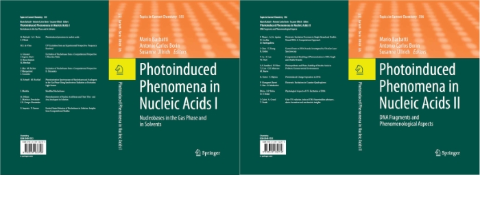
Topics in Current Chemistry

Photoinduced Phenomena in Nucleic Acids

M. Barbatti, A. C. Borin, S. Ullrich: Photoinduced Phenomena in Nucleic Acids I, Top. Curr. Chem. 355, Springer 2015

M. Barbatti, A. C. Borin, S. Ullrich: Photoinduced Phenomena in Nucleic Acids II, Top. Curr. Chem. 356, Springer 2015


Photoinduced processes in nucleic acids are phenomena of fundamental interest in diverse fields, from prebiotic studies, through medical research on carcinogenesis, to the development of bioorganic photodevices. In this contribution we survey many aspects of the research across the boundaries. Starting from a historical background, where the main milestones are identified, we review the main findings of the physical-chemical research of photoinduced processes on several types of nucleic-acid fragments, from monomers to duplexes. We also discuss a number of different issues which are still under debate.

C. Z. Bisgaard, H. Satzger, S. Ullrich, A. Stolow: Excited state dynamics of isolated DNA bases: A case study of Adenine, ChemPhysChem 2009, 10, 101.

M. D. Lee, J. D. Coe, S. Ullrich, M.-L. Ho, S.-J. Lee, B.-M. Cheng, M. Z. Zgierski, I-C. Chen, T. J. Martínez, A. Stolow: Substituent effects on Dynamics at Conical Intersections: alpha,beta –enones, J. Phys. Chem. A 2007, 111, 11948.

H. R. Hudock, B. G. Levine, A. L. Thompson, H. Satzger, D. Townsend, N. Gador, S. Ullrich, A. Stolow, T. J. Martínez: Ab Initio Molecular Dynamics and Time-Resolved Photoelectron Spectroscopy of Electronically Excited Uracil and Thymine, J. Phys. Chem. 2007, 111, 8500.

H. Satzger, D. Townsend, M. Z. Zgierski, S. Patchkovskii, S. Ullrich, A. Stolow: Primary processes underlying the photostability of isolated DNA bases: adenine,PNAS 2006,103, 10196.

E.Samoylova, H. Lippert, S. Ullrich, I.V. Hertel, W. Radloff, T. Schultz: Dynamics of photoinduced processes in adenine and thymine base pairs, J. Amer. Chem. Soc. 2005, 127, 1782.

S. Ullrich: Investigation of biomolecular photostability by fs time-resolved photoelectron spectroscopy, LabPlus International (October 2004 issue).

S. Ullrich, T. Schultz, M.Z. Zgierski, A. Stolow: Electronic relaxation mechanism in DNA and RNA bases studied by time-resolved photoelectron spectroscopy, Phys. Chem. Chem. Phys. 2004, 6, 2796. Invited article for special issue on "Bio-active Molecules in the Gas-phase".

S. Ullrich, T. Schultz, M.Z. Zgierski, A. Stolow: Direct observation of electronic relaxation dynamics in adenine via time-resolved photoelectron spectroscopy, J. Amer. Chem. Soc., Communication 2004, 126, 2262.

T. Schultz, S. Ullrich, J. Quenneville, T. J. Martinez, M. Z. Zgierski, A. Stolow: Azobenzene photoisomerization: Two states and two relaxation pathways explain the violation of Kasha's rule. in: Femtochemistry and Femtobiology: Ultrafast Events in Molecular Science, M. M. Martin, J. T. Hynes eds. (Elsevier, Amsterdam 2004) pp. 45-48.

M. Smits, C.A. deLange, S. Ullrich, T. Schultz, M. Schmitt, J.G. Underwood, J.P. Shaffer, D.M. Rayner, A. Stolow: Stable kilohertz-rate molecular beam laser ablation sources, Rev. Sci. Instrum. 2003, 74, 1.

S. Ullrich, K. Müller-Dethlefs: A REMPI and ZEKE spectroscopic study of trans-acetanilide•H2O and comparison to ab initio CASSCF calculations J. Phys. Chem. A 2002, 106, 9188.

S. Ullrich, K. Müller-Dethlefs: A REMPI and ZEKE spectroscopic study of a secondary amid group in Acetanilide, J. Phys. Chem. A 2002, 106, 9181.

X. Tong, S. Ullrich, C.E.H. Dessent, K. Müller-Dethlefs: Intermolecular vibration and internal rotation of a methyl group in acetanilide•Ar: ZEKE photoelectron spectroscopy study, Phys. Chem. Chem. Phys. 2002, 4, 3578.

S. Ullrich, G. Tarczay, X. Tong, K. Müller-Dethlefs: Hydration of a Cationic Amide group: A ZEKE Spectroscopic Study of the trans-Formanilide•H2O Complex, Phys. Chem. Chem. Phys. 2002, 4, 2897.

S. Ullrich, G. Tarczay, X. Tong, M.S. Ford, C.E.H. Dessent, K. Müller-Dethlefs: A REMPI and ZEKE Spectroscopic Study of the trans-Formanilide•Ar van der Waals Cluster, Chem. Phys. Lett. 2002, 351, 121.

S. Ullrich, G. Tarczay, X. Tong, C.E.H. Dessent, K. Müller-Dethlefs: ZEKE Photoelectron Spectroscopy of the Cis- and Trans-Isomers of Formanilide, Ang. Chem. Int. Ed., Communication 2002, 41, 166.

S. Ullrich, G. Tarczay, K. Müller-Dethlefs: A REMPI and ZEKE spectroscopic study of the Phenol×Kr and Phenol•Xe van der Waals complexes., J. Phys. Chem. A 2002, 106, 1496.

S. Ullrich, G. Tarczay, X. Tong, C.E.H. Dessent, K. Müller-Dethlefs: A ZEKE Photoelectron Spectroscopy and Ab Initio Study of the Cis- and Trans-Isomers of Formanilide: Characterizing the Cationic Amide Bond?, Phys. Chem. Chem. Phys. 2001, 3, 5450.

W.D. Geppert, S. Ullrich, C.E.H. Dessent, K. Müller-Dethlefs: Observation of Rotational Isomers II: A ZEKE and Hole-Burning Spectroscopy Study of Hydrogen-Bonded 3-Methoxyphenol•H2O Clusters, J. Phys. Chem. A 2000, 104, 11870.

S. Ullrich, W.D. Geppert, C.E.H. Dessent, K. Müller-Dethlefs: Observation of Rotational Isomers I: A ZEKE and Hole-Burning Spectroscopy Study of 3-Methoxyphenol, J. Phys. Chem. A 2000, 104, 11864.

C.E.H. Dessent, W.D. Geppert, S. Ullrich, K. Müller-Dethlefs: Ionization-induced conformational changes: REMPI and ZEKE spectroscopy of salicyl and benzyl alcohol, Chem. Phys. Lett. 2000, 319, 375.

W.D. Geppert, C.E.H. Dessent, S. Ullrich, K. Müller-Dethlefs: Observation of Hydrogen-Bonded Rotational Isomers of the Resorcinol•Water Complex, J. Phys. Chem. A 1999,103, 7186.

R.F. Delmdahl, S. Ullrich, K.-H. Gericke: Photofragmentation of OClO(Ã2A2ν1ν2ν3)→Cl(2PJ)+O2, J. Phys. Chem. A 1998, 102, 7680.